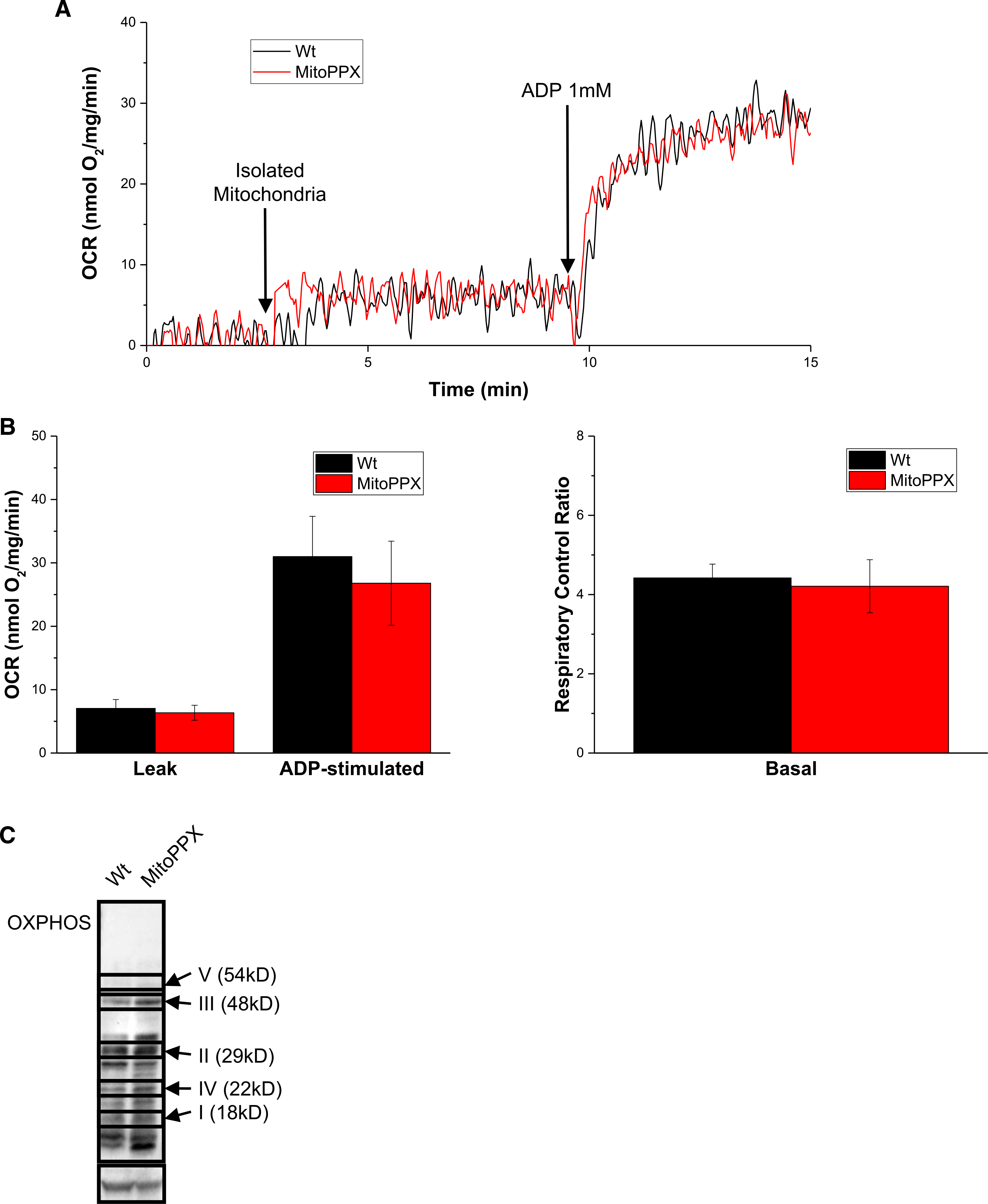
Figure 4. Isolated mitochondria from MitoPPX cells show Wt activity.

(A) OCR measurements of mitochondria isolated from either Wt or MItoPPX cells. (B) Analysis of OCR and the respiratory control rate of Wt and MitoPPX cells during the respirometry experiment. (C) Steady-state levels of different components of the respiratory chain in from Wt and MitoPPX HEK 293 cells, using Western blotting. Immunoblot was conducted using a commercially available cocktail antibody, containing all the components of the ETC. Bands not labeled are unspecific. Actin was used as loading control. Data in the graphs are shown as average ± SEM.
