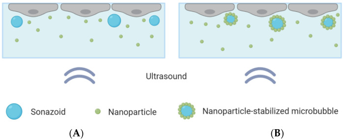
Figure 1.
Schematic overview of the CLINIcell with a cell layer on top, microbubbles and nanoparticles in solution and ultrasound applied from below. (A): Cells exposed to nanoparticles co-incubated with Sonazoid. (B): Cells exposed to nanoparticle-loaded microbubbles and an excess of nanoparticles.

