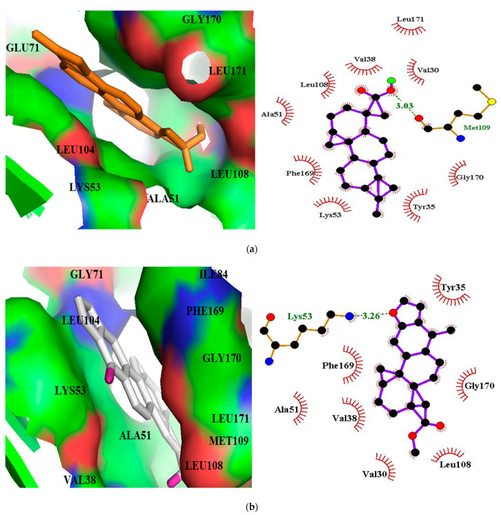
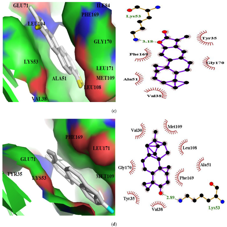
Figure 3.
Docking poses and protein-ligand interaction studies of top four hits with the lowest binding energies (a) ZINC95486106, (b) ZINC95913720, (c) ZINC33832090, and (d) ZINC95919076 against p38 MAPK structure. The binding pockets are represented as surfaces and the ligands as sticks. In the LigPlot+ representations, the ligands are displayed as purple sticks, hydrophobic contacts are shown as red spoke arcs, and the hydrogen bonds with their respective bond lengths as green.


