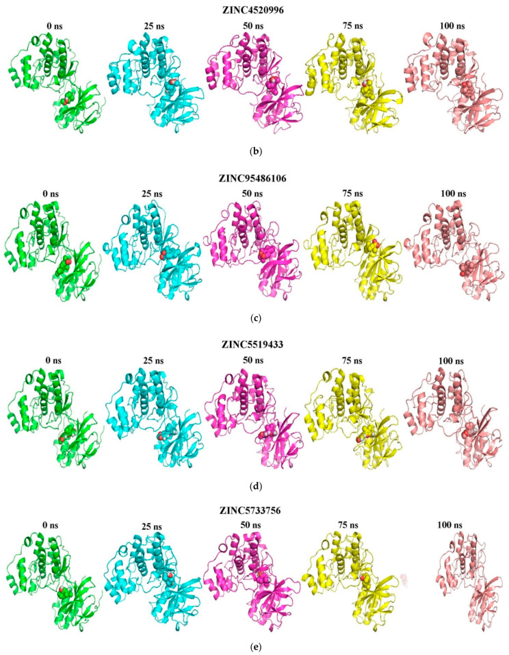
Figure 5.
Snapshots at 25 ns intervals ((time step = 0, 25, 50, 75, and 100 ns) for the binding modes of the ligand-p38 MAPK complexes. The cartoon representation shows (a) ZINC1691180, (b) ZINC4520996, (c) ZINC95486106, (d) ZINC5519433 and (e) ZINC5733756- p38 MAPK complexes. The ligands are represented as spheres and the protein as cartoons. All the ligands were observed to bind stably in the ATP binding pocket.


