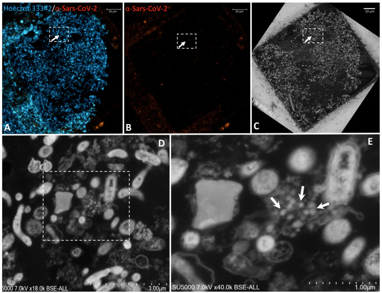
Figure 3.
Correlative light fluorescence and electron microscopy in an ultra-thin section of sewage sample. Confocal laser scanning microscopy images of 100-nm-thick ultra-thin section (Z maximal projection) of a sewage sample (A,B), DNA stained with Hoechst 33342 (blue) and the SARS-CoV-2 particles labelled with anti-SARS-CoV-2 spike protein (orange-red). Scanning electron microscopy images (C–E) of the ultra-thin section shown in (A,B). The boxed region of interest in ((A–C), scale bars 10 µm) is shown at a higher magnification in ((D), scale bar 3 µm). Boxed region in (D) is zoomed in ((E), scale bar 1 µm) with hypo-electron dense circular structures surrounded by a hyper-dense crown-like shapes with 75–100-nm diameters (arrows) are present in the boxed region positive for anti-SARS-CoV-2 fluorescence.

