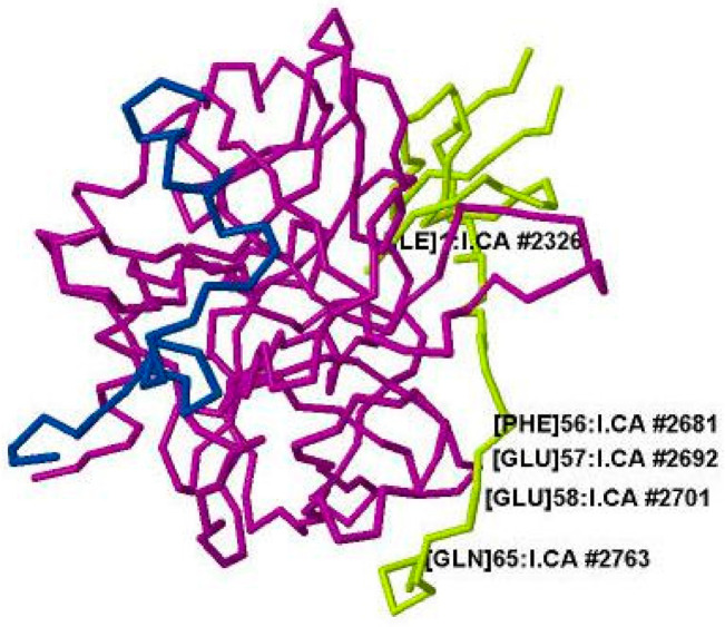
Figure 12.
The strand structures of the A chain in blue and B chain in cyan of human-α -thrombin complexed with hirudin in light green. Ile1′ and the most likely site of a short hydrogen bond (SHB) occurring in the thrombin-r-hirudin complex, residues 55 to 65 of r-hirudin engaged with Arg75 and Arg77 of thrombin, are labeled. The image is from the Protein Data Bank PDB file 4HTC [129] modeled with Jmol Version 12.0.41.

