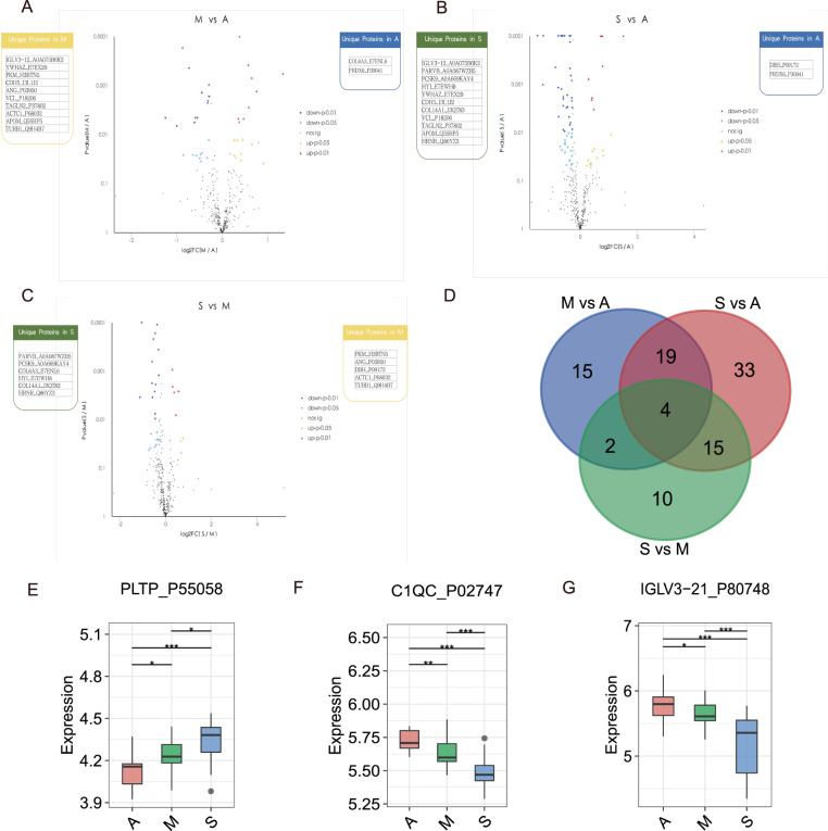
Fig. 6. DEPs identification among the diverse recovered patients groups.
Volcano plots of DEPs in M vs A (A), S vs A (B), and S vs M (C) treatments. The x-axis presents the log2FC and the y-axis presents −log10 (p value). The warm color triangles indicate significantly upregulated genes (red: p-value < 0.01; yellow: p-value < 0.05). The blue inverted triangles indicate downregulated genes (navy: p-value < 0.01; lightblue: p-value < 0.05), and black points indicate non-significantly different genes. The blue, yellow, and green boxes on either side of each volcano plot represent unique proteins in A, M, and S groups, successively. D Venn plot for identified DEPs among the diverse recovered patients groups. The blue, red, and green circles represent M vs A, S vs A, and S vs M groups, respectively. Box plots for PLTP_P55058 (E), C1QC_P02747 (F), IGLV3-21_P80748 (G) expression levels in A, M, and S treatments.

