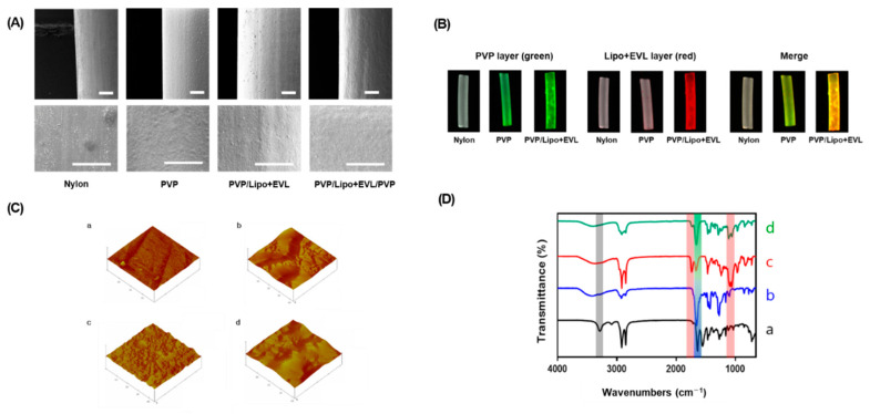
Figure 4.
Characterization of PVP, PVP/Lipo + EVL, and PVP/Lipo + EVL/PVP-coated surface of Nylon tubes. (A) SEM images of the coated surface (Scale bars equal to 400 μm). (B) Fluorescence images of the coated surface using FOBI. GreenL 5(6)-Carboxyfluorescein-stained PVP, Red: Nile red-labeled liposome. (C) AFM images for surface roughness evaluation. (D) ATR-FTIR analysis for Nylon tube surface coated with PVP, PVP/Lipo + EVL, and PVP/Lipo + PVP/PVP. (a) Bare Nylon tube, (b) PVP-coated Nylon tube, (c) PVP/Lipo + EVL-coated Nylon tube, and (d) PVP/Lipo + EVL/PVP-coated Nylon tube.

