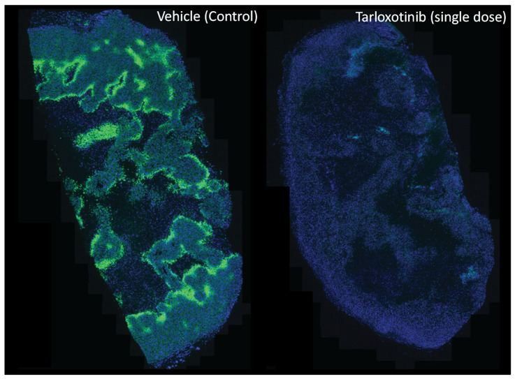
Figure 4.
Modification of the hypoxic microenvironment following administration of tarloxotinib in PC9 mutant-EGFR NSCLC tumour-bearing mice. The representative immunohistochemical image shows hypoxic regions (green, pimonidazole binding) across a tumour cross section before and after (72 h) a single dose of tarloxotinib (30 mg/kg, intraperitoneal). Nuclei are counterstained (blue, DAPI). Image was provided by Dr Shevan Silva with permission.

