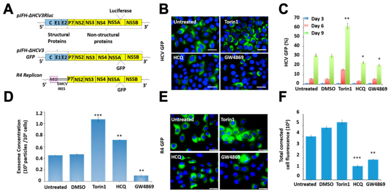
Figure 1.
Illustrated is the impact of autophagy induction, lysosomal degradation, and exosome release on virus-cell survival in persistently infected HCV culture. (A) Schematic of the construct used for HCV infection and the sub-genomic replicon cell line. The impact of autophagy induction, lysosome inhibitor (HCQ), and exosome inhibitor (GW4869) on HCV GFP expression by flow analysis. Huh-7.5 cells were infected at the HCV GFP virus at MOI 0.01. Infected cells on days 3, 6 and 9 were treated with 2 rounds of Torin1 (100 nM), HCQ (10 μM), or GW4869 (10 μM). (B) Fluorescence image of HCV-GFP expression. (C) Quantification of HCV-GFP by flow analysis at 3, 6 and 9 days after treatments. (D) Nanoparticle tracking analysis (NTA) shows the impact of Torin1, HCQ and GW4869 treatment on extracellular vesicle release in R4GFP replicon cells. (E) Fluorescent microscopy images of untreated R4GFP replicon cells and treated with Torin1 (100 nm), HCQ (10 μM) and GW4869 (10 μM) for 72 h. (F). Fluorescence intensity measurements representing the GFP expression of replicon cell culture. * p < 0.05, ** p < 0.01, *** p < 0.001. Error bars represent the standard deviation (SD) of 3 measurements.

