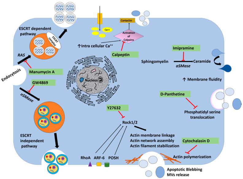
Figure 8.
The action mechanism of drugs used to inhibit extracellular vesicle release. Extracellular vesicles originate from the endosomal pathway or pinch off from the cell membrane. Exosomes are produced from MVBs by ESCRT-dependent and ESCRT-independent pathways. While manumycin A inhibits exosome dependent pathway, GW4869 inhibits exosome independent pathway. Microvesicle biogenesis is modulated by lipids and cytoskeletal proteins. Lipid rafts and cholesterol play an important role in the budding of cell membranes. Enzymes involved in the transfer of lipid from one leaflet of the cell membrane to the other are potentially targeted to inhibit exosome release. Calpeptin is a family of calcium-dependent neutral cytosolic cysteine protease inhibitors used as a micro-vesicular inhibitor. Y27632 is a competitive inhibitor of both ROCK1 and ROCK2 and is able to compete with ATP in binding to the catalytic site of these kinases. This compound inhibits microvesicle release by blocking these two proteins. This compound reorganizes the cytoskeleton and mediates cellular contractility by regulating the activity of actin filaments. Imipramine is a well-known antidepressant that promotes membrane fluidity by inhibiting acid sphingomyelinase (aSMase), therefore preventing the generation of microvesicles. D-Pantethine inhibits cholesterol synthesis as well as fatty acid synthesis as the fluidity of the cell membrane is important during membrane bi-layer reorganization and microvesicle formation. This drug blocks the translocation of phosphatidylserine to the outer surface membrane, which is an essential step of microvesicle formation. Cytochalasin D is an alkaloid produced as a toxin by many fungi. This compound binds the edges of actin filaments to prevent actin polymerization. Actin polymerization is essential for the formation of membrane-derived microvesicles and their intracellular movement.

