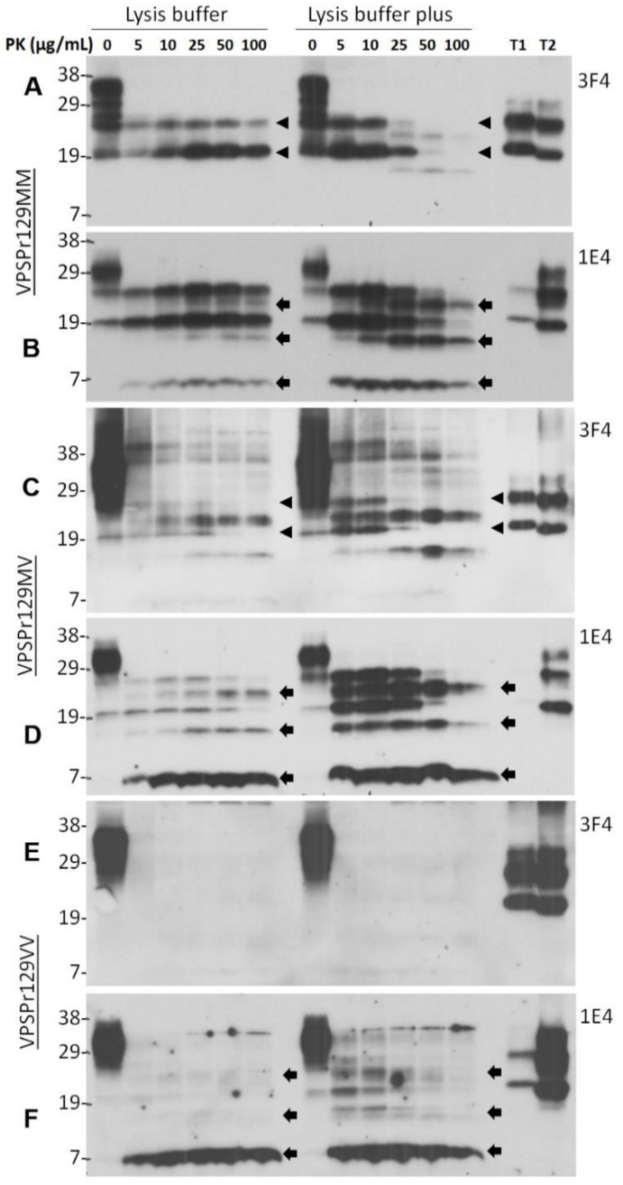
Figure 1.
PK concentration-dependent two-step fragmentation of PrPSc from VPSPr. Brain homogenates from cadavers of VPSPr129MM (A,B), VPSPr129MV (C,D), and VPSPr129VV (E,F), homogenized in standard lysis buffer (pH 7.4) (left sides of western blots) or in lysis buffer “plus” (pH 8.0) (right sides of western blots) were treated with PK at different concentrations ranging from 0, 5, 10, 25, 50, to 100 µg/mL prior to western blotting with 3F4 (A,C,E) or 1E4 (B,D,F). T1: PrPSc sCJD type 1 control; T2: PrPSc sCJD type 2 control. Arrow heads indicate ~26 kDa and ~20 kDa PrPres fragments while arrows indicate ~23 kDa, ~17 kDa and ~7 kDa PrPres fragments, respectively.

