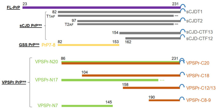
Figure 8.
Schematic diagram of various PK-resistant PrPSc in VPSPr, sCJD and GSS. Purple rectangle represents the full-length (FL)-PrP23–231 with the GPI anchor (blue circular arrow). Gray rectangles: sCJDT1: sCJD PrPSc type 1 with an N-terminal starting residue 82 [45]; sCJDT2: sCJD PrPSc type 2 with an N-terminal starting residue 97 [45]; sCJD-CTF13: sCJD PrPSc C-terminal fragment with an N-terminal starting residue 154 [9]; sCJD-CTF12: sCJD PrPSc C-terminal fragment with an N-terminal starting residue 162 [9]. T1AF: Anchorless fragment of PrPSc type 1 with an unknown C-terminal truncation site (…) [46]; T2AF: Anchorless fragment of PrPSc type 2 with an unknown C-terminal truncation site (…) [46]. Yellow rectangle: GSS PrPSc fragment PrP7–8 with a N-terminal starting residue 82 and a C-terminal ending residue 153 from [43]. Green rectangles represent the 1E4-detected VPSPr PrPSc fragments with anticipated N-terminal starting at residue 86, but different C-termini [14] (current study): VPSPr-N20 with an intact GPI anchor, VPSPr-N17 with a molecular weight of 17 kDa but an unknown C-terminal truncation site (…), and VPSPr-N7 with an anticipated C-terminal ending residue 145. Red rectangles represent the anti-C antibody-detected VPSPr PrPres fragments with anticipated intact C-termini carrying GPI anchor [14] (current study): VPSPr-C20 with an N-terminal starting residue 86, VPSPr-C18 with an anticipated N-terminal truncation site at residue 104, VPSPr-C12/13 with an anticipated N-terminal truncation site at residue 158, and VPSPr-C8–9 with an anticipated N-terminal truncation site at residue 190.

