Figure 2.
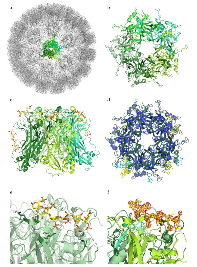
Glycan engagement of HPV16. (a) Capsid structure of HPV16 L1 pentamers (PDB ID 5KEP). L1 subunits of one pentamer are displayed in different shades of green. The highlighted pentamer is represented as a ribbon, the remaining capsid is depicted as cartoons. (b) Top view of a HPV16 L1 pentamer (PDB ID 5W1O). Subunits are displayed in cartoon representation and colored in different shades of green. (c) Side view of the pentamer. The heparin ligand is colored by atom type and represented with sticks in yellow. (d) Superposition of HPV16 L1 pentamer in green with its heparin ligand in yellow with HPV18 L1 pentamer (PDB ID 5W1X) in blue and its ligand in cyan. (e) Close-up view of one ligand binding site of HPV16. Ligand-binding residues K356, T358, K361, and K54 are represented as sticks. (f) Close-up view as in (e) of one ligand binding site with the negative Fo–Fc difference map of the ligand contoured at 3σ, colored in red. Figures were created using PyMOL [23].

