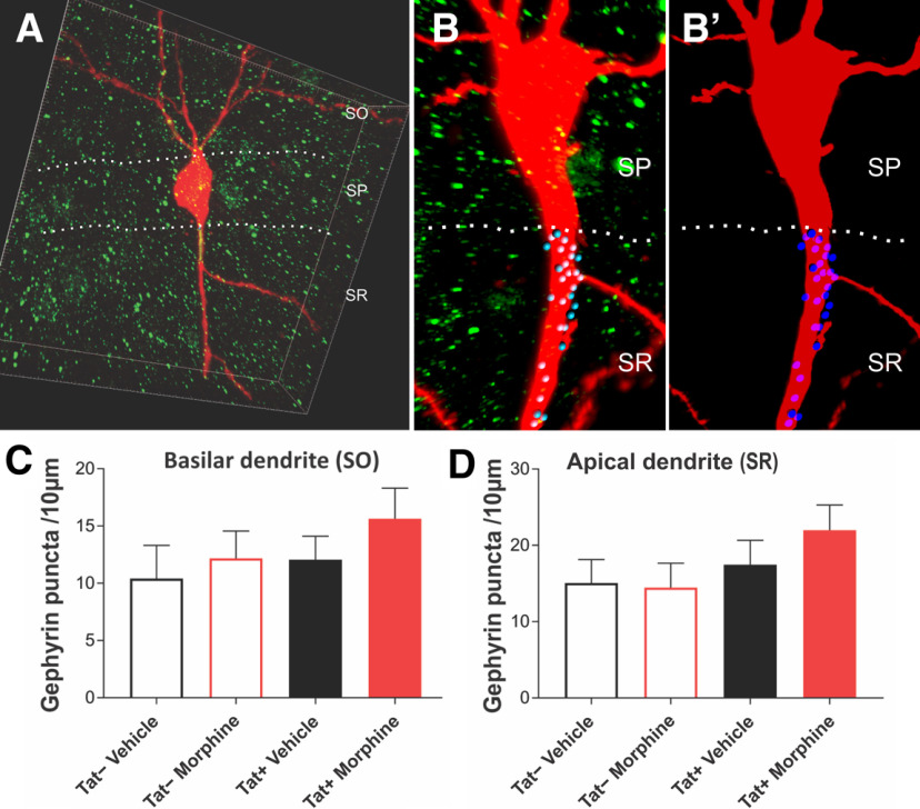
Figure 6.
Assessment of inhibitory, postsynaptic gephyrin puncta within the aspinous, proximal dendrites of CA1 pyramidal cells. Biocytin-filled pyramidal cells labeled with an Alexa Fluor 594-conjugated streptavidin probe (red) and gephyrin puncta (green) were analyzed. A, B, B’, A third channel (blue, with darker blue puncta are in front of the dendrite; lighter blue puncta are behind the dendrite; B, B’), only identifying the gephyrin puncta that were adjacent or overlapped with the aspinous portions of the basilar (C) or perisomatic apical (D) pyramidal cell dendrite, from 3D-reconstructed images was quantified. No changes in the number of gephyrin-immunoreactive puncta were observed. Box Dimensions in A: 100 × 100 × 20 μm. Data represent the mean number of gephyrin-positive puncta ± SEM per 10-μm length of dendrite.

