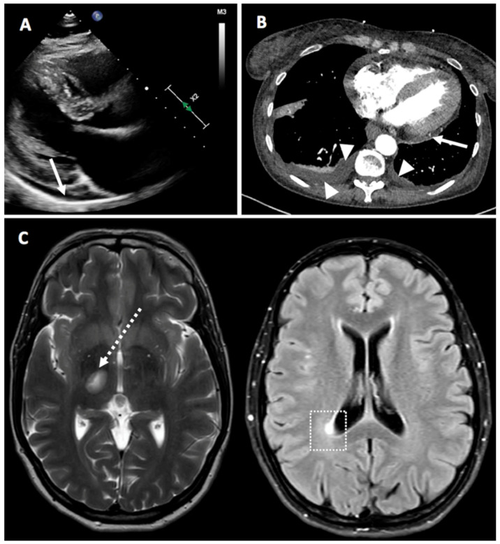
Figure 4.
Panel (A) (Case 4)—Parasternal, long axis view from a 2-D echocardiogram showing a small-sized posterior pericardial effusion (white arrow). Panel (B) (Case 27)—Computerized tomography scan demonstrating posterior pericardial effusion (white arrow) and bilateral pleural effusion (white arrowheads). Panel (C) (Case 7)—Magnetic resonance imaging of the brain showing demyelinating lesions in the left mesencephalic (dotted white arrow) and in the right peri-ventricular occipital white matter (white dotted square).

