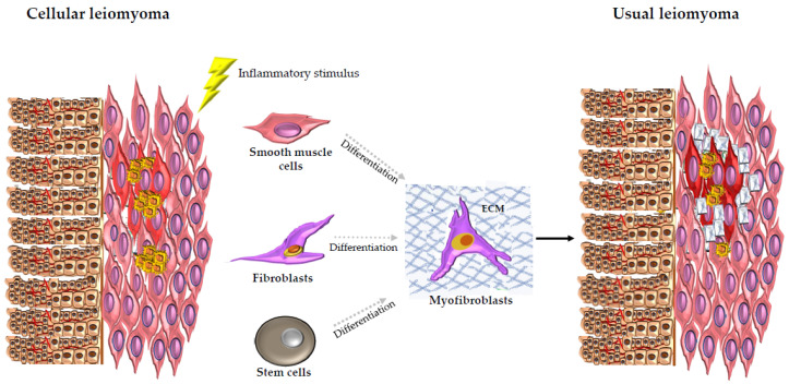
Figure 5.
Illustration of the possible phase mechanism of leiomyoma development proposed by our group. Cellular leiomyoma is considered as the first step in the tumoral transformation. In fact, cellular leiomyoma shows higher levels of macrophage (yellow in the figure) infiltration and an increased number of inflammatory cells. This aspect could represent a response to an inflammatory stimulus that leads some cellular leiomyoma cells to myofibroblast differentiation with the consequent upregulation of the typical extracellular matrix (ECM) proteins. In fact, usual leiomyoma shows a larger amount of ECM proteins and low levels of macrophage infiltration. So, usual leiomyoma could be considered as the late-phase tumor. The blue net represents the typical ECM proteins: collagen 1A1, fibronectin and versican. The red color represents the uterine fibroids (light red for cellular leiomyoma histotype and dark red for usual leiomyoma histotype). The pink color represents the myometrium; the brown color represents the endometrium. The blood vessels within endometrium are also represented (red lines in the figure).

