Table 1.
Different phantoms used for MPI reconstruction.
| Year | Journal | Phantoms | Description |
|---|---|---|---|
| 2016 | IEEE Transactions on Medical Imaging |
|
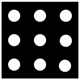
The size of the cube-shaped calibration sample size is 2 × 2 × 2 mm3. The calibration sample is moved in vertical and horizontal steps of 2 mm over the 30 × 30 mm2 FOV [6]. |
| 2017 | IEEE Transactions on Medical Imaging |
|
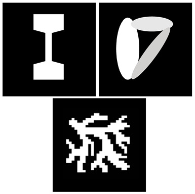
Three different phantoms with one percent Gaussian noise are used to evaluate the reconstruction quality: a simulated stenosis, overlapping ellipses, and a vascular tree [45]. |
| 2018 | Journal of Mathematical Imaging and Vision |

|
The first is a typical resolution phantom with round objects of different size and concentration. The second includes three ellipses with different size and concentration. The third simulates a situation where objects cannot be covered by a single FOV [62]. |
| 2019 | IEEE Transactions on Medical Imaging |
|
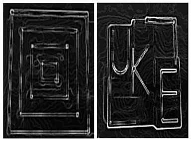
The first is the filled 3D-printed model, which consists of four rectangles with different sizes. The second is the UKE phantom. The letters of the phantom are located in different planes in the y direction [38]. |
| 2019 | Physics in Medicine & Biology |
|
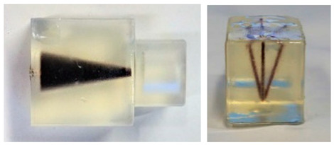
These two phantoms are from the open MPI datasets (www.tuhh.de/ibi/research/open-mpi-data.html (accessed on 7 October 2020)). The first is a cone and the second consists of five tubes with a common origin on one side of the phantom [56]. |
| 2019 | Measurement |
|
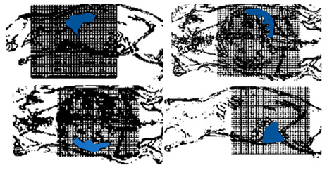
Real images are used to study MPI reconstruction, which represent the different mouse organs: the lungs, left kidney, right kidney, and reproductive system [7]. |
