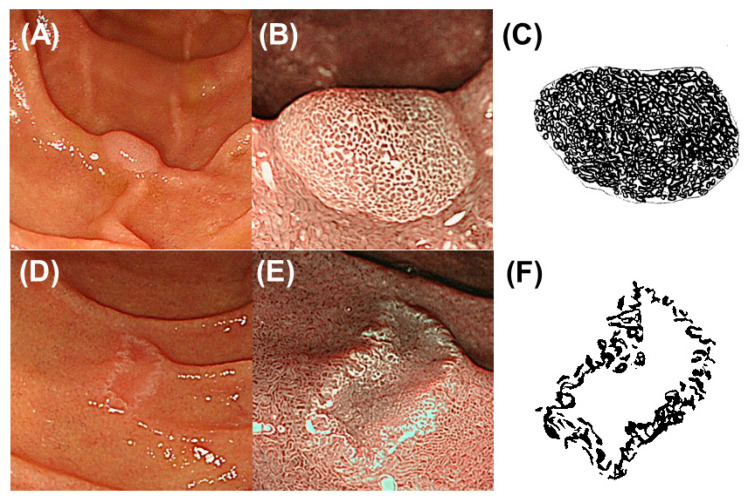
Figure 1.
Representative images of entire-type and non-entire-type milk-white mucosa (MWM). (A,B) Representative endoscopic images of a superficial non-ampullary duodenal epithelial tumor (SNADET) with entire-type MWM. (A) White-light endoscopic image showing a small, elevated lesion. (B) Narrow band imaging (NBI) magnification endoscopic image showing MWM throughout the elevated lesion corresponding to entire-type MWM. (C) Schematic diagram of entire-type MWM. The black area shows the endoscopic MWM-positive area throughout the lesion. The endoscopic MWM-positive rate was 95%. (D,E) Representative endoscopic images of a SNADET with non-entire type MWM. (D) White-light endoscopic image showing a small, depressed lesion. (E) NBI magnification endoscopic image showing MWM only along or near the margin corresponding to non-entire type MWM. (F) Schematic diagram of non-entire type MWM. The endoscopic MWM-positive rate was 40%.

