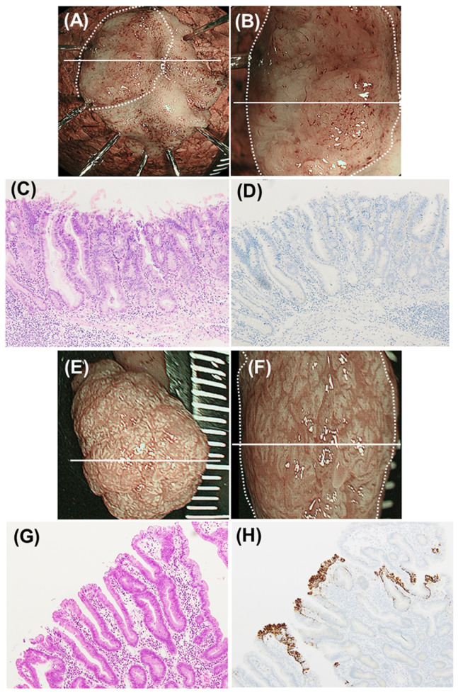
Figure 2.
Representative images of milk-white mucosa (MWM)-positive and -negative superficial non-ampullary duodenal epithelial tumor (SNADET) lesions. (A–D) Representative images of a SNADET of high-grade intraepithelial neoplasia (HGIN). (A) Narrow band imaging (NBI) endoscopic image of an endoscopically resected specimen. The white line indicates the maximal section. The endoscopic MWM-positive rate was 0% (non-entire type). (B) NBI magnification endoscopic image of the resected specimen. The white line indicates the maximal section. (C) Histology of the maximal section (hematoxylin-eosin (HE) staining). (D) Immunohistochemical finding of the maximal section (adipose differentiation-related protein (ADRP) staining). Neoplastic epithelial cells were all negative for ADRP staining. (E–H) Representative images of a SNADET of low-grade intraepithelial neoplasia (LGIN). (E) NBI endoscopic image of an endoscopically resected specimen. The white line indicates the maximal section. The endoscopic MWM-positive rate was 95% (entire type). (F) NBI magnification endoscopic image of the resected specimen. The white line indicates the maximal section. (G) Histology of the maximal section (HE staining). (H) Immunohistochemical finding of the maximal section (ADRP staining). Most neoplastic epithelial cells (90%) were positive for ADRP staining.

