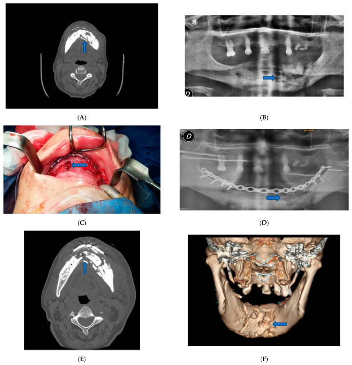
Figure 1.
Patient 1. (A) Pre-operative CT scan image showed lithic area in the mandibular horizontal branches, which appeared subverted and fractured on the left side. (B) Pre-operative panoramic X-ray image of the dental branch revealed lithic area. (C) Epi-mucosal fixation performed using SMART Lock screws and plates without elevating the mucoperiosteal flap. (D) Post-operative panoramic X-ray image of the dental branch. (E) Post-operative CT scan image. (F) Post-operative CT scan 3D reconstruction.

