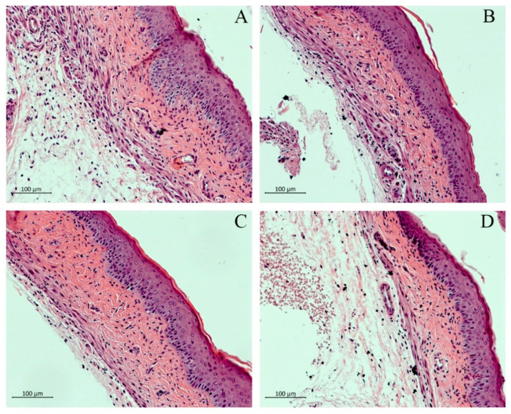
Figure 3.
Histopathological analysis of vaginal tissues with H & E staining. Mouse vaginal tissue was excised longitudinally, fixed in 10% neutral-buffered formalin, and then embedded in paraffin. Each section of paraffin-embedded tissues was stained with H & E and then observed using alight microscope. (A) Naive group, (B) Blank, (C) CAR (50 mg/kg), and (D) WSCP1 (50 mg/kg). Magnification: 400×; scale bars, 100 μm.

