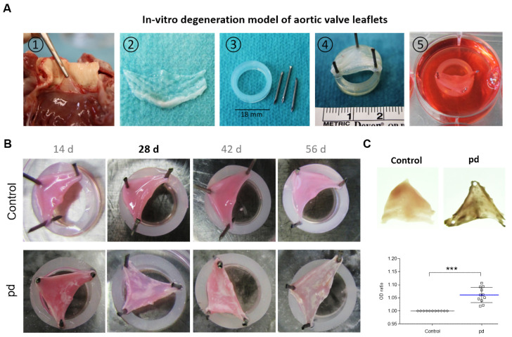
Figure 1.
In vitro degeneration model of aortic valve leaflets. (A) Application of in vitro CAVD model: (1) Preparation of AV leaflets. (2) Excised AV leaflets after washing in PBS. (3) Required materials (silicon rubber rings and needles). (4) AV leaflet stretched on silicon rubber ring. (5) Cultivation of tensed AV leaflets. (B) Images of temporal progression of AV leaflet degeneration. White areas indicate calcified domains. (C) Representative transmitted light images of AV leaflets after 28 d cultivation and analysis of optical density (OD). Data (n = 8) are mean ± SEM. p-values are calculated by using Student’s t-test with Dunn’s multiple comparison post hoc test.; ***: p < 0.001. Pd, (pro-degenerative) condition.

