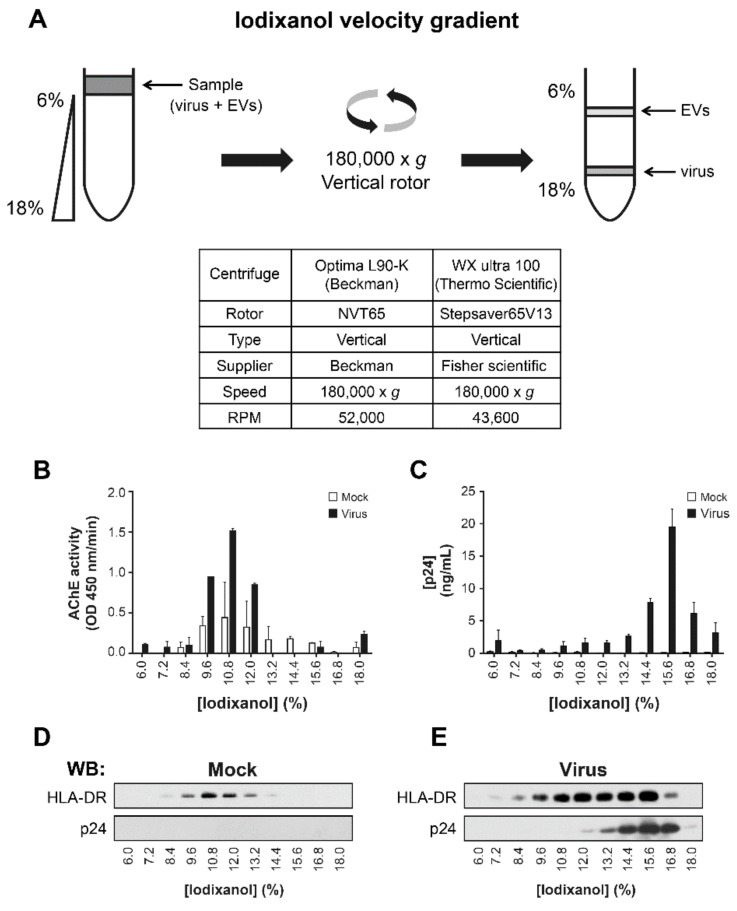
Figure 1.
Iodixanol velocity gradient separates HIV-1 from EVs in infected cell-lines. In a continuous 6–18% iodixanol gradient (OptiprepTM), the sample is laid on the top of the gradient and ultracentrifuged in a vertical rotor for 50–75 min (A). Raji-CD4 cells were cultured with NL4-3 virus or mock for five days. Microfiltered supernatant was centrifuged at 100,000× g for 45 min. The pellet was re-suspended in PBS and laid on an iodixanol velocity gradient and centrifuged for 50 min. The relative abundance of EVs based on AChE activity (B) and the abundance of the virus based on p24 ELISA (C) were assessed. EVs from Raji-CD4 cells were recovered as described above and laid on iodixanol velocity gradient and centrifuged for 75 min. Precipitated proteins were probed for host membrane protein such as HLA-DR or viral protein p24 by western blots (D,E).

