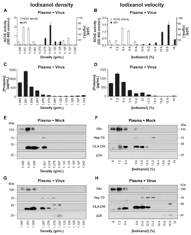
Figure 5.
Separation of plasma proteins, EVs, and HIV-1 in the iodixanol density and velocity gradients. A pool from five plasma samples from HIV-1 negative individuals were spiked with virions or mock from Raji-CD4 infected-cells. Then EVs were precipitated by ExoQuickTM. The pellet was re-suspended in PBS and laid on iodixanol density (left panels) or velocity (right panels) gradients. Acetylcholinesterase (AChE) (A,B), p24 (A,B), total (C,D), and specific proteins (E–H) were quantified using an enzymatic assay, an ELISA, BCA kit, and western blot respectively.

