Figure 1.
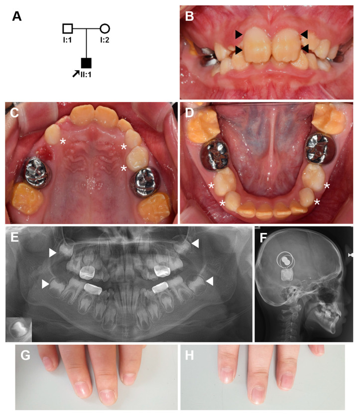
Pedigree, clinical photos, and radiographs of the proband. (A) Pedigree of the family. The arrow indicates the proband, and individual IDs are shown below the symbols. (B–D) Frontal, maxillary and mandibular clinical photos of the proband at age 8 years 7 months. The buccal side of the maxillary central incisors shows relatively normal looking areas in the middle and cervical thirds (black arrow heads). Deciduous teeth (white asterisks) do not have hypoplastic enamel or discoloration, but the permanent teeth show hypoplastic enamel and brown discoloration. (E) A panoramic radiograph revealed hypoplastic enamel in the developing permanent dentition. Almost no covering enamel can be easily seen in the second molars (white arrow heads) compared to the normal tooth in an inset at the lower left corner. (F) Bilateral cochlear implants can be seen in the lateral cephalogram. (G,H) His finger nails are normal.

