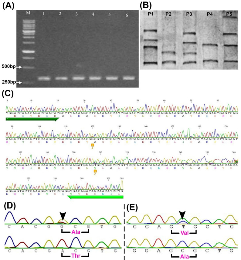
Figure 1.
Identification of SNPs in buffalo PRLR(L2). (A) Agarose gel of PRLR(L2) amplified fragments (305 bp) from six different samples (lanes 1-6). (B) PCR-SSCP banding patterns show five different patterns (P1-P5) in five different samples. (C) A representative sequence chromatogram from one sample shows the sites of the two SNPs (two orange boxes), primers (forward (F) and reverse (R), the two green arrows), and amino acid sequences (colored letters). (D,E) Sequences chromatogram spanning the site of g.11685G>A (D) and g.11773T>C (E) SNPs (arrowheads) and the altered amino acids (p.Ala494Thr and p.Val523Ala). Ala, alanine; M; 1Kb ladder; Thr, threonine; Val, valine.

