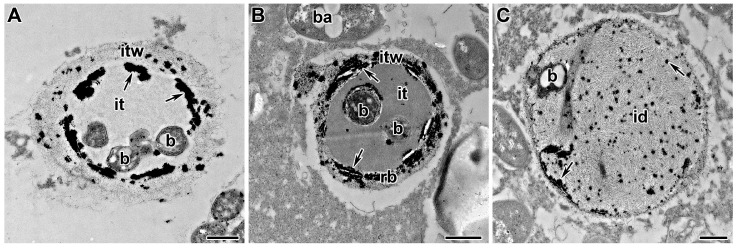
Figure 4.
Sequential steps of hardening of the infection thread wall and matrix during development and growth in the wild-type nodules of Pisum sativum. (A) Localization of H2O2 in the inner side of infection thread wall. Incomplete hardening of the wall of the infection thread makes it possible to form an infection droplet and release the Rhizobium into the plant cell. (B) Localization of H2O2 across the entire infection thread wall thickness. Complete hardening of the infection thread wall prevents the formation of an infection droplet and the release of bacteria. (C) Localization of H2O2 inside infection droplet matrix. Hardening of the infection droplet prevents the growth and division of bacteria inside the lumen. Cytochemical localization of H2O2 as electron-dense precipitate formed in the presence of cerium chloride. id—infection droplet, it—infection thread, itw—infection thread wall, b—bacterium. Arrows indicate electron-dense precipitates. Bars = 500 nm.

