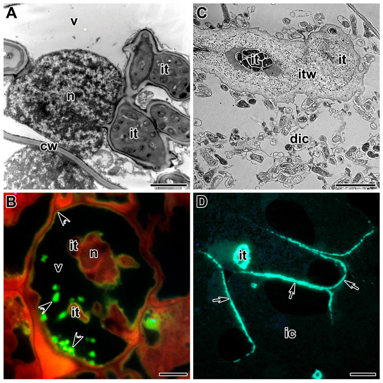
Figure 5.
Defense reactions in the nodules of symbiotic Pisum sativum mutants. (A) Transmission electron micrograph of abnormal infection threads in the nodule of mutant SGEFix–-2 (Pssym33-3). (B) Suberin depositions in the infection thread walls and around vacuole in the nodule of mutant SGEFix–-2 (Pssym33-3), which is characterized with ‘locked’ infection threads. Neutral red staining for detection of suberin. (C) Transmission electron micrograph of an abnormal infection thread in the nodule of mutant RisFixV (Pssym42). (D) Callose (β-1,3-glucan) depositions in the infection thread wall and cell wall of infected cells in the nodule of mutant RisFixV (Pssym42), which is characterized with abnormal infection threads and early senescence of symbiotic structures. Callose depositions detected by staining with Aniline blue. ic—infected cell, dic—degenerated infected cell, n—nucleus, v—vacuole, cw—cell wall, it—infection thread, itw—infection thread wall. Arrows indicate callose depositions in cell walls of infected cells, arrowheads indicate suberin depositions in the vacuole. Bars (A) = 2 µm, (B–D) = 5 µm.

