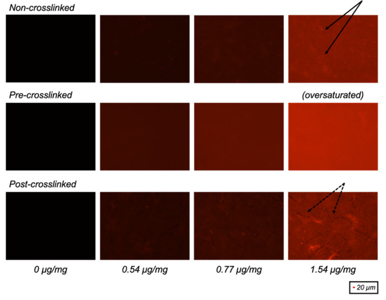
Figure 1.
Fluorescence images of non-crosslinked, pre-crosslinked and post-crosslinked collagen films with different quantities of 78% dye-conjugated dendrimer activation (µg dendrimer per mg collagen). Presence of overexposed bright particulates (solid) and underexposed collagen “coils” (dashed) are indicated by black arrows.

