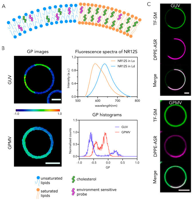
Figure 2.
Membrane order in Lo and Ld phases of synthetic (GUVs) and cell-derived (GPMVs) membranes: (A,B) Visualization of phase separation in synthetic and cell-derived membrane systems using phase sensitive probes; (A) Cartoon of phase separation and distribution of phase- sensitive probes; (B) Ratiometric imaging of phase separation in GUVs and GPMVs using the environmental-sensitive probe NR12S. Fluorescence spectrum of NR12S exhibits a drastic red shift in Ld in comparison to Lo. The images of membranes at 560 nm and 650 nm emission wavelengths were recorded and used to produce GP images. The color code on the GP images corresponds to the color bar: dark red is +1, and dark blue is −1. The GP histograms for both GUV and GPMV show two distinct populations. In each case, low GP counts correspond to Ld whereas high GP counts correspond to Lo. Importantly, GP values of Ld and Lo are different for GUVs and GPMVs. (C) TopFluor-labeled SM (TF-SM, green) partitions into Ld in GUVs (DOPC:SM:Chol, 2:2:1) while it partitions to both phases in GPMVs derived from U2OS cells. The DPPE-Abberior Star Red (DPPE, ASR, magenta) partitions preferentially to Ld in both GUVs and GPMVs. Scale bars are 5 µm.

