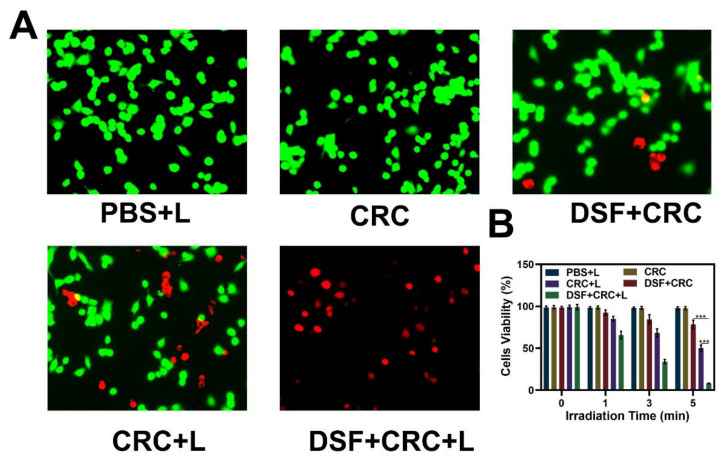
Figure 3.
(A) Fluorescence images of 4T1 cells stained with fluorescein diacetate (FDA) (live cells, green fluorescence) and propidium iodide (PI) (dead cells, red fluorescence) after incubation with different formulations. (B) Cell viability of 4T1 cells cultured in the presence of various formulations after laser irradiation. The p-values were calculated from a Tukey post-test, where *** p < 0.001.

