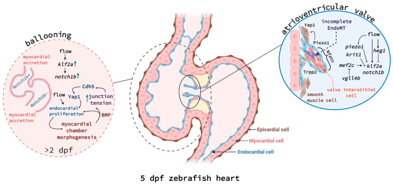
Figure 1.
Endocardium-myocardium signalling during ballooning (left) and atrioventricular valve formation (right). (Left) Cell communication between endocardial and myocardial cells is critical for chamber morphogenesis and is also linked to blood flow. Endocardial cells in atrium and ventricle show distinct expression patterns, e.g., ventricular endocardial cells express much higher levels of Notch1b protein. Endocardial cells are proliferating in response to flow and induce myocardial expansion. Myocardial cells do not proliferate during ballooning but rather increase in cell size and by accretion at the poles. Bmp produced by myocardial cells and the increasing junctional tension, mediated by Cadherin-5 (Cdh5) and Yap1, in turn increase endocardial cell proliferation, creating a feed-forward loop. (Right) A subset of endocardial cells undergoes incomplete EndoMT to fold into cardiac valves. Tissue tension, mediated by Piezo1 and Trpp2, increases Yap1 expression in some endocardial and surrounding smooth muscle cells to help the elongation of the valve leaflet. Endocardial cells at the tip of the valve are expressing high levels of klf2a and notch1b in response to flow, but a network of signalling pathways linked to krit1, heg1, vgll4b, and piezo1 causes downregulation of klf2a in endocardial cells closer to the myocardium and cardiac jelly. Interstitial cell differentiation and proliferation is dependent on transcription factor Nfatc. Figure created with Biorender.com.

