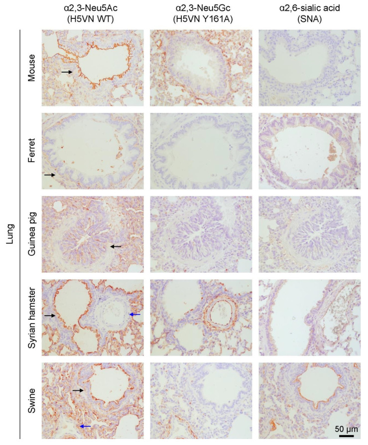
Figure 2.
Visualization of the presence of α2,3 linked Neu5Ac (WT HA of A/Vietnam/1203/2004 (H5N1)), α2,3 linked Neu5Gc (Y161A mutant HA of A/Vietnam/1203/2004 (H5N1)), and α2,6 linked sialic acids (SNA) using AEC staining on lung tissue of mouse (C57BL/6), ferret, guinea pig, Syrian hamster, and domestic swine. Brown staining indicates binding of the HAs to the tissue and blue indicates the cells. Selected bronchioles (black arrows) and blood vessels (blue arrows) are indicated. Images are representative of two independent experiments.

