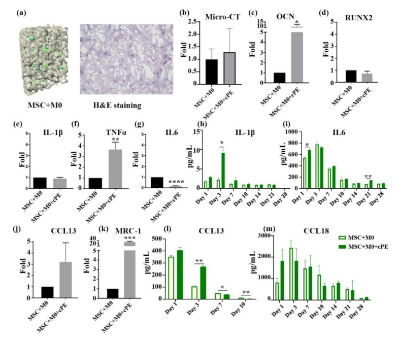
Figure 2.
Effects of M0 macrophages on MSC osteogenesis in cPE environment. M0 macrophages polarized to M1 and M2 macrophages and enhanced the osteogenesis of MSCs in the cPE treatment group. (a) Cross-sectional illustration of the 3D co-culture. MSCs were co-cultured with M0 macrophages with or without cPE. H&E staining shows cells evenly distributed throughout the scaffold. (b) Micro-CT analysis of scaffolds. The amount of mineralization did not differ significantly in the cPE group. (c,d) qPCR data of osteogenic gene expression. (e–g) qPCR result of inflammation related genes. (h,i) Levels of secreted pro-inflammatory cytokines. IL-1β and IL6 were detected. (j,k) mRNA expressions of CCL13 and MRC were determined by qPCR. (l,m) Cytokine profile of CCL13 and CCL18 in the medium. (* p < 0.05, ** p < 0.01, *** p < 0.001).

