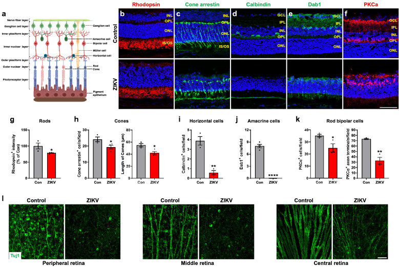
Fig. 2.
ZIKV infection impairs retinal neurons. a Schematic illustration of retinal structure (generated using BioRender). b–f Immunohistochemical staining on retinal sections from control and ZIKV-infected mice with retinal specific markers, including rhodopsin for rods, cone arrestin for cones, calbindin for horizontal cells, Dab1 for amacrine cells, PKCα for rod bipolar cells at P21. Blue: DAPI staining for nuclei. g–k Graphs represent the quantification of these cells. l Representative images of Tuj1-stained RGCs and their axons in the peripheral, middle and central area of retinal flatmounts at P21. Scale bar = 50 μm. Data are presented as mean ± S.E.M. n = 3–4 per group. *p < 0.05, **p < 0.01, ****p < 0.0001 compared to control mice

