
Figure 3:
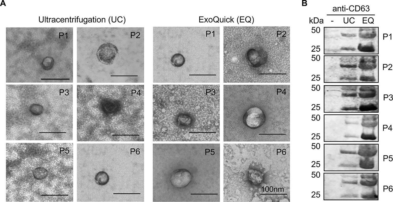
Validation of exosome enrichment from human cell-free sera. (A) TEM micrographs of exosomes in ultracentrifugation (UC) and ExoQuick (EQ) preparations. Data for 6 independent patient samples are shown (P1–6). Exosomes confirmed by size (30–100nm) and appearance. Scale bar in each image represents 100 nm. (B) Immunoblot of CD63 in unprocessed cell-free serum alone (−), UC, and EQ exosome preparations. Adapted from Prendergast et al. (2018)124, Copyright @ 2018, PLOS.