
Figure 8.
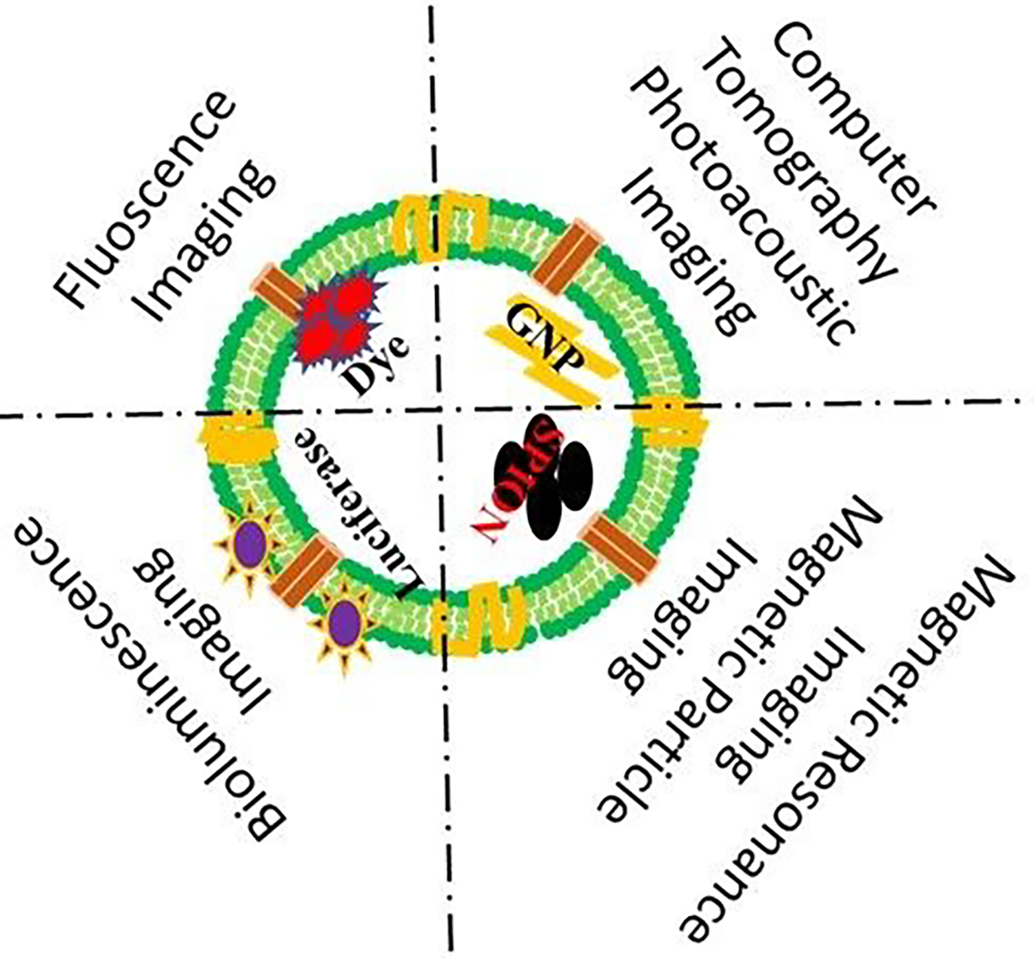
Exosome visualization using various imaging modalities. Fluorescence dye-labeled or luciferase-expressing exosomes visualize the biodistribution or tissue uptake under optical imaging systems (fluorescence imaging or bioluminescence imaging). Gold nanoparticles (GNPs) labeling exosomes observe the whole-body tracking in deep tissues under computer tomography (CT) or photoacoustic imaging (PAI). Superparamagnetic iron oxide nanoparticles (SPIONs) labeled exosomes show the active cell migration or homing to target regions in vivo under magnetic resonance imaging (MRI) or magnetic particle imaging (MPI).