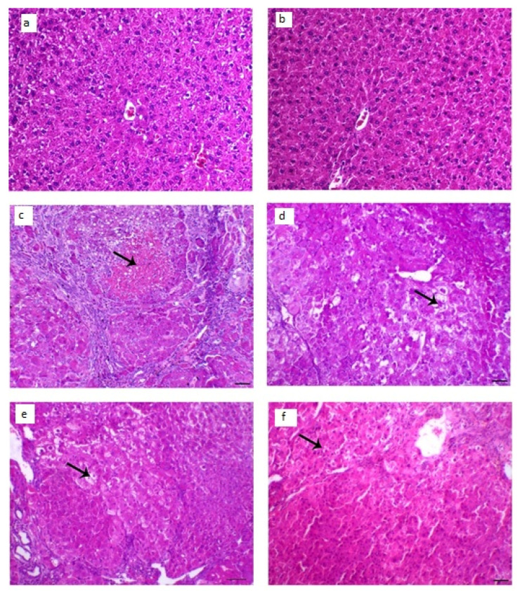
Figure 10.
Histopathological changes in liver tissues; (a,b) liver of control animal appearing typical hepatocytes organized in lines around the central vein (arrow; H&E staining; scale bar, 100 µm, (c,d) liver section of the diseased animal showing HCC nodule revealing marked hepatic necrosis (arrow), H&E, bar = 100 µm., (e) liver segment of infected animal treated with LUT-suspension showing hepatic adenoma with hepatic vacuolar degenerative changes (arrow), H&E, bar = 100 µm, (f) liver segment of infected animal treated with LUT-ENPs appearing hepatic foci with marked diminish the hepatic neoplastic injuries with a little number of hepatic adenomas (arrow shows central degenerative zone), H&E, bar = 100 µm.

