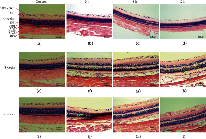
Figure 4.

Histopathological features of the retina in each group. (a–d) The structure and morphology of the retina in the 4-week control group and daily exposure of 3 h, 6 h, and 12 h experimental groups. There was no significant change in the IS/OS and RPE layer structure of the retina in the 3 h, 6 h, and 12 h experimental groups. (e–h) The structure and morphology of the retina in the 8-week control group and 3 h, 6 h, and 12 h experimental groups. The IS/OS layer was loose and edematous (green arrow) at 3 h, 6 h, and 12 h after 8 weeks with thinning of the RPE layer and the decrease of the number of cells. (i–l) The structure and morphology of the retina in the 12-week normal control group and 3 h, 6 h, and 12 h experimental groups. The IS/OS layer was loose and edematous (green arrow) lightly stained and swollen (green arrow) with thinning of the RPE layer and the decrease of the number of cells. The scale bar is 50 μm. These tests were repeated 3 times for each rat, and there were 6 rats per group. NFL: nerve fiber layer; GCL: ganglion cell layer; IPL: inner plexiform layer; INL: inner nuclear layer; OPL: outer plexiform layer; ONL: outer nuclear layer; IS/OS: photoreceptor inner/outer segment layers; RPE: retina pigmented epithelium.
