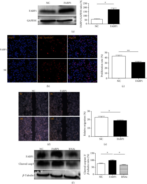
Figure 3.

FABP1 inhibits the proliferation and migration of liver cancer cells and promotes apoptosis. (a) Western blot results of FABP1 overexpression in Huh-7 cells. Huh-7 cells were transfected with the FABP1 lentivirus (FABP1), and the blank lentivirus (NC) was used as control. Quantification of the Western blot (right panel). Data are expressed as the means ± SD, n = 3 in each group. ∗P < 0.05. (b) Huh-7 cell proliferation after FABP1 overexpression. The nuclear was stained by DAPI in blue; the proliferative cells were stained by EdU-Apollo567 in red. 10 randomly selected fields were checked under a fluorescence microscope. (c) Quantification of the proliferation rate (%). The proliferation ratio was calculated as the number of proliferating cells/total number of cells × 100% by ImageJ software. Data are expressed as the means ± SD, n = 3 in each group. ∗∗P < 0.01. (d) Huh-7 cell migration showed by the scratch assay after FABP1 overexpression; 10 randomly selected fields were checked under an inverted microscope. (e) Quantification of the migration rates (%). Data are expressed as the means ± SD, n = 3 in each group. ∗P < 0.05. (f) Huh-7 cell apoptosis after FABP1 overexpression or knockdown. The FABP1 overexpression or RNAi lentivirus was used for infection. Quantification of Western blot was shown in the right panel. Data are expressed as the means ± SD, n = 3 in each group. ∗P < 0.05.
