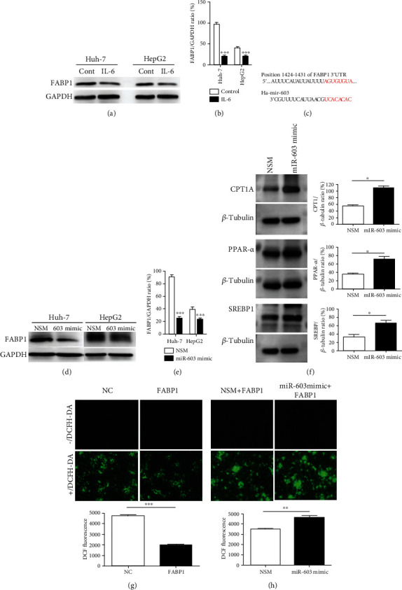
Figure 5.

FABP1 reduces ROS levels in HCC cells, while miR-603 reverses the inhibitory effect of FABP1 on oxygen free radicals. (a) Western blot results of FABP1 expression level after IL-6 treatment in Huh-7 and HepG2 cells. (b) Quantification of the Western blot. Data are expressed as the means ± SD, n = 3 in each group. ∗∗∗P < 0.001. (c) The binding sites of FABP1 mRNA 3′-UTR (1424 to 1431) to miR-603 were shown as red. (d) Western blot results of FABP1 expression level after miR-603 treatment in Huh-7 and HepG2 cells. (e) Quantification of the Western blot. Data are expressed as the means ± SD, n = 3 in each group. ∗∗∗P < 0.001. (f) Western blot results of lipid metabolism- and synthesis-related protein expression after miR-603 mimic transfection in Huh-7 cells. Quantification of the Western blot was shown in the right panel. Data are expressed as the means ± SD, n = 3 in each group. ∗P < 0.05. (g) ROS levels after FABP1 overexpression treatment. Cells transfected with the FABP1 overexpression lentivirus or NC were stained with DCFH-DA (green), and 10 randomly selected fields were checked under a fluorescence microscope. Quantification of the DCF fluorescence was shown in down panel. Data are expressed as the means ± SD, n = 3 in each group. ∗∗∗P < 0.001. (h) ROS levels after miR-603 treatment. Cells were transfected with the FABP1 lentivirus or NC. Next, the cells were transfected with miR-603 (FABP1+miR-603 mimic) or NSM (FABP1+NSM) and stained with DCFH-DA; 10 randomly selected fields were checked under a fluorescence microscope. Quantification of the DCF fluorescence was shown in down panel. Data are expressed as the means ± SD, n = 3 in each group. ∗∗P < 0.01.
