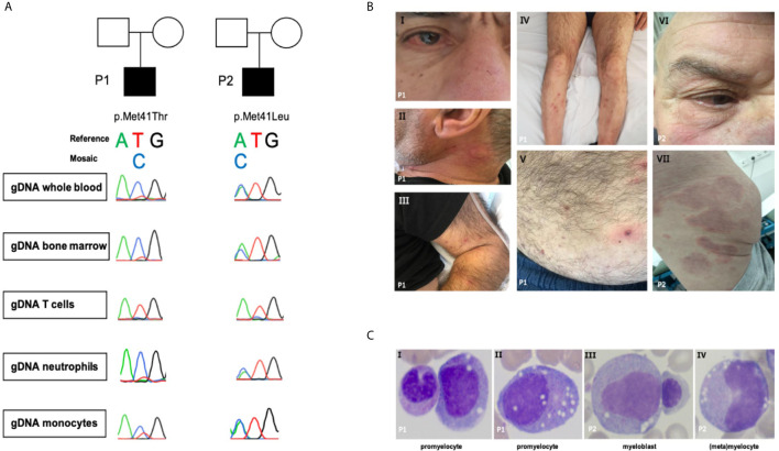
Figure 1.
(A) Sanger sequence of UBA1. (B.I) Episcleritis, nose chondritis. (B.II) Urticarial lesion of the neck. (B.III-V) Nodular lesions on extremities and abdomen: biopsy-confirmed complement-mediated vasculopathy. (B.VI) Episcleritis, periorbital erythema (B.VII) Urticarial vasculitis of gluteal region. (C) Representative aberrant vacuolized bone marrow cells (x40 magnification); left panel also a normal band neutrophil, third panel also a normal erythroblast.

