Figure 5.
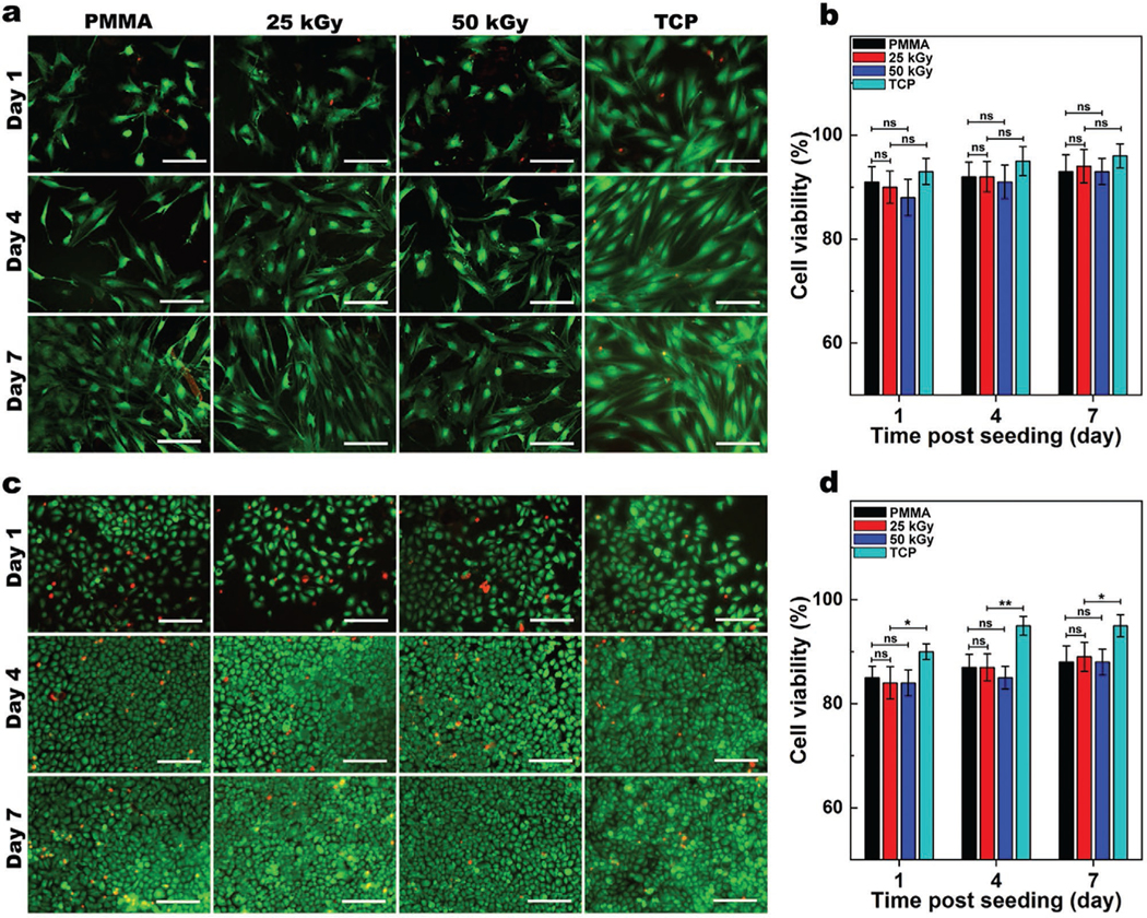
Biocompatibility of PMMA with and without E-beam irradiation. Representative Live/Dead images of human corneal fibroblasts (HCF) a) and human corneal epithelial (HCEp) cells c) cultured on PMMA, with and without 25 or 50 kGy irradiation compared to those cultured on tissue culture plates (TCP), and their corresponding cell viability b,d) after 1, 4, and 7 days of cell culture (scale bar: 200 μm). Cells viability was quantified from Live/Dead images using ImageJ (Green [calcein AM]: lived cells; Red [ethidium homodimer-1]: dead cells). Values are presented as mean ± SD; n = 4. ns, *, **, ***, and **** represent p > 0.05, p ≤ 0.05, p ≤ 0.01, p ≤ 0.001 and p ≤ 0.0001.

