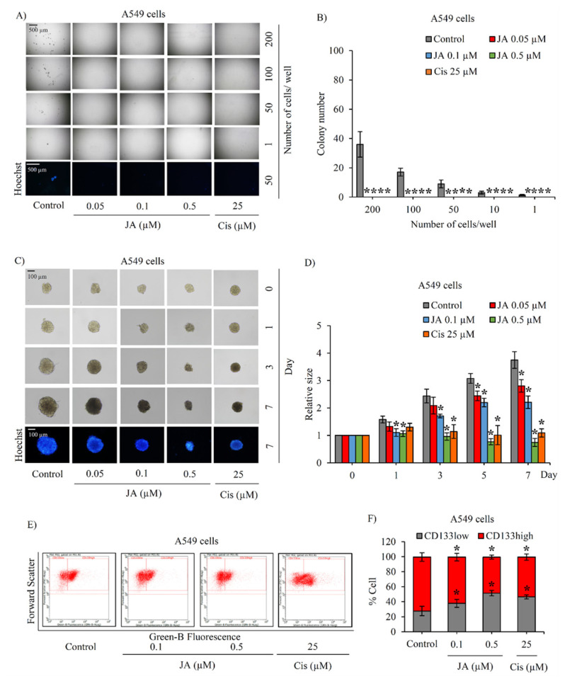
Figure 5.
Suppressive effect of jorunnamycin A on stem-like phenotypes of lung cancer A549 cells. (A) Jorunnamycin A (JA) inhibited capability to generate cancer spheroid of human lung cancer A549 cells in limiting dilution assay (LDA) for 14 days at all concentrations. Optical microscopy (4×) was used to capture spheroid images, while fluorescence microscopy (10×) was used to take images with blue fluorescence from Hoechst33342 staining. (B) The number of forming colonies at all cell densities used was decreased in all groups exposed to nontoxic concentrations (0.05–0.5 µM) of JA. (C) Suppressive effect of JA in CSC-enriched A549 lung cancer cells was evaluated by single three-dimensional (3D) CSC spheroid formation. The enlargement of CSC spheroids was suppressed by JA (0.05–0.5 µM). At day 7 of treatment, all single 3D CSCs were photographed by optical microscopy (10×) and spheroid images depicting bright blue fluorescence of Hoechst33342 were obtained by fluorescence microscopy (10×). (D) Culture with JA (0.1–0.5 µM) illustrated the significant reduction of relative spheroid size of CSC-enriched lung cancer cells at days 1–7. (E) Flow cytometry analysis of CD133 expression confirmed the reduction of CD133high population after treatment with JA (0.1–0.5 µM) for 3 days. (F) JA at 0.1–0.5 µM diminished CD133high population in CSC spheroids compared with untreated control. Cisplatin (Cis) at 25 µM was used as a positive control. The colony number (B) and relative size (D) were respectively analyzed from cancer colonies (A) and CSC-enriched spheroids (C). The %cell demonstrated in (F) was calculated from dot plots presented in (E). Data represent means ± SD of three independent experiments. * p < 0.05 versus non-treated control.

