Figure 7.
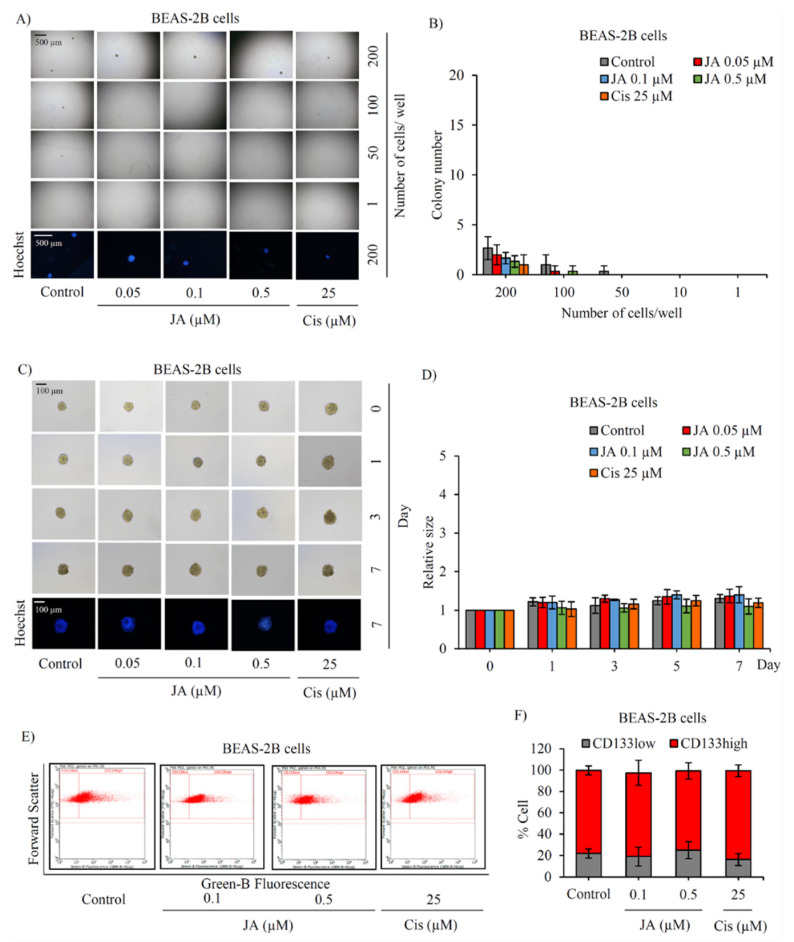
No alteration of stemness phenotypes in human lung epithelial cells cultured with jorunnamycin A. (A) Limiting dilution assay (LDA) revealed a low amount of colony formation obtained from normal lung epithelial BEAS-2B cells that were cultured in ultra-low attachment 96-well plate for 14 days. (B) There was no significant difference in the number of colonies generated from BEAS-2B cells incubated with 0.05–0.5 µM jorunnamycin A (JA), compared with untreated cells. Optical microscopy (4×) was used to capture spheroid images, while fluorescence microscopy (10×) was used to take images with blue fluorescence from Hoechst33342 staining. (C) Stemness features of BEAS-2B cells were also examined in single three-dimensional (3D) spheroid formation. After culture for 7 days, all single 3D spheroids were photographed by optical microscopy (10×) and spheroid images depicting bright blue fluorescence of Hoechst33342 were obtained by fluorescence microscopy (10×). Neither (D) relative spheroid size nor (E,F) %CD133-overexpressing cells in BEAS-2B spheroids were altered after treatment with JA (0.1–0.5 µM) when compared with untreated control. Cisplatin (Cis) at 25 µM was used as a positive control. The colony number (B) and relative size (D) were respectively analyzed from forming colonies (A) and stem cell-enriched spheroids (C). The %cell demonstrated in (F) was calculated from dot plots of flow cytometry analysis as presented in (E). Data represent means ± SD of three independent experiments.

