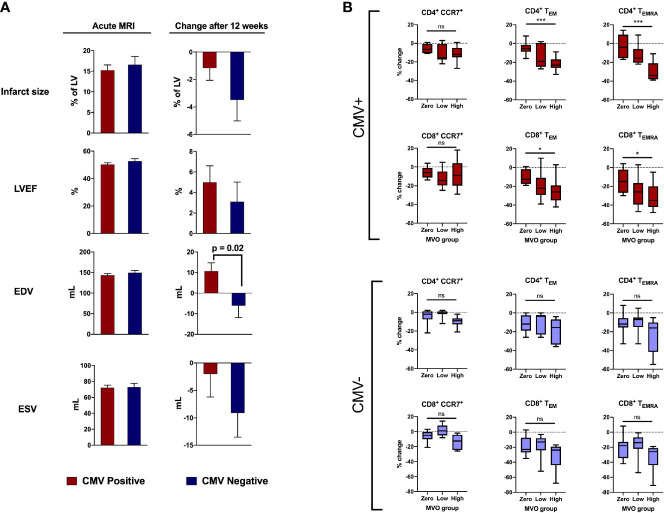
Figure 2.
CMV serostatus is associated with adverse remodeling 12 weeks after reperfusion. (A) In the acute phase (1-8 days post-reperfusion, n=101), CMV serostatus has no effect on infarct size, LV ejection fraction, end-diastolic volume or end-systolic volume. At 12-week follow-up (n=48), CMV seropositive patients displayed significantly more deterioration in end-diastolic volume (+10.7mL vs -6.1mL, p=0.02). P values determined using the unpaired t-test. (B) Relationship between amount of MVO (zero, low or high) and change in T cell subsets between 15-30 minutes post-reperfusion, separately for CMV seropositive and seronegative patients. Box plots display median (central line), 25th and 75th centiles (limits of box), and range (error bars). Statistics refer to differences between MVO groups as indicated (Kruskal-Wallis test with Dunn’s multiple comparisons test). Total n=47; CMV positive n=25 [8 zero MVO, 6 low, 11 high], CMV negative n=22 [9 zero MVO, 7 low, 6 high]). * p<0.05, ***p<0.001; ns, not significant.

