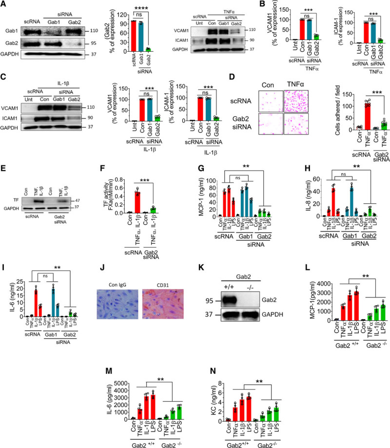
Figure 1.

Gab2 (Grb2-associated binder2) silencing or deficiency inhibits TNFα (tumor necrosis factor alpha)-induced, IL (interleukin)-1β–induced, or lipopolysaccharide (LPS)-induced proinflammatory responses in endothelial cells. A, Human umbilical vein endothelial cells (HUVECs) were transfected with 200 nmol/L scrambled siRNA (scRNA), Gab1 (Grb2-associated binder 1), or Gab2 siRNA. After 48 h, the transfected cells were analyzed for the expression of Gab1 and Gab2 proteins by immunoblot analysis. Band intensities were quantified by densitometry, and these data are shown in the right. B and C, Gab1 or Gab2 silenced cells were treated with TNFα (10 ng/mL; B) or IL-1β (10 ng/mL; C) for 6 h. The cell lysates were analyzed for VCAM1 (vascular cell adhesion molecule 1) and ICAM1 (intercellular adhesion molecule 1) protein levels by immunoblot analysis. The blots shown on the left were representative. Band intensities were quantified by densitometry, and these data are shown in the right. D, HUVECs transfected with scrambled siRNA or Gab2 siRNA were treated with TNFα (10 ng/mL) for 6 h and then incubated with THP1 monocytic cells (5×10⁵/mL). After 30 min, the nonadherent THP-1 cells were removed, and HUVEC monolayers were washed with serum-free medium. The adherent cells were fixed with 4% paraformaldehyde at room temperature for 30 min. The cells were stained using crystal violet dye. The adherent cells were visualized under a bright-field microscope at ×20 magnification. The images in the left depict representative images. The number of cells adhered/field was determined by counting multiple fields in ≥3 experiments performed independently and averaging them per field (right). E and F, HUVECs transfected with scrambled siRNA or Gab2 siRNA were treated with TNFα and IL-1β (10 ng/mL each) for 6 h, and TF (tissue factor) expression was analyzed by immunoblotting (E) or functional activity (F). TF functional activity was measured by adding FVIIa (10 nmol/L) and substrate factor X (175 nmol/L) to the cells in a calcium-containing buffer and measuring the amount of FXa generated at 5 min. G–I, HUVECs were transfected with scrambled, Gab1, or Gab2 siRNA. After 48 h, the transfected cells were treated with TNFα (10 ng/mL), IL-1β (10 ng/mL), or LPS (500 ng/mL) for 15 h. The supernatants were collected, and the levels of MCP1 (macrophage chemoattractant protein 1; G), IL-8 (H), and IL-6 (I) were measured in ELISA. J, Brain endothelial cells isolated from Gab2−/− mice were stained with endothelial marker CD31 (cluster of differentiation). K, Immunoblot showing the complete absence of Gab2 in the brain endothelial cells from Gab2−/− mice. L and M, Mouse brain endothelial cells isolated from Gab2−/− or WT (wild type) littermate control mice were serum starved overnight and treated with TNFα (20 ng/mL), IL-1β (20 ng/mL), or LPS (1 µg/mL) for 15 h. The supernatants were collected and assayed for MCP-1 (L), IL-6 (M), and KC (keratinocyte-derived chemokine, the mouse ortholog of human interleukin-8; N) levels in ELISA. For data shown in A through D, Student t test was used to calculate statistically significant differences. For others, 1-way ANOVA was used to compare the data of experimental groups, and Tukey post hoc multiple comparison test was used to obtain statistical significance between the two groups. ns indicates no statistically significant difference. **P<0.01, ***P<0.001.
