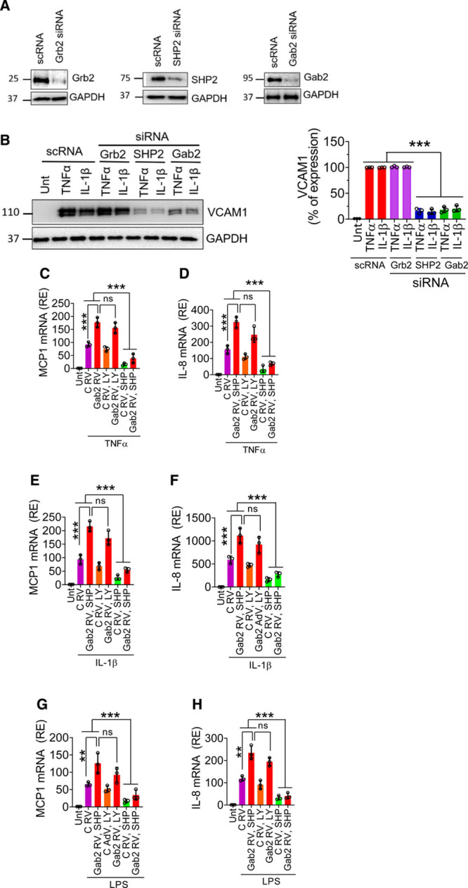
Figure 2.

The involvement of Grb2 (growth factor receptor-bound protein 2), PI3K (phosphoinositide 3-kinase), or SHP2 (SH2 containing protein tyrosine phosphatase-2) in Gab2 (Grb2-associated binder2)-mediated inflammatory signaling. A, Human umbilical vein endothelial cells (HUVECs) were transfected with 200 nmol/L scrambled, Grb2, SHP2, or Gab2 siRNA for 48 h. The gene silencing was confirmed by immunoblotting. B, The transfected cells were treated with TNFα (tumor necrosis factor alpha), IL (interleukin)-1β (10 ng/mL), or lipopolysaccharide (LPS; 500 ng/mL) for 6 h. VCAM1 (vascular cell adhesion molecule 1) expression was analyzed by immunoblot analysis. Band intensities were quantified by densitometry, and the quantified data are shown in the right. C–H, HUVECs were left untreated (Unt), infected with control (C RV) or Gab2 retrovirus (Gab2 RV) were treated with LY294002 (5 µmol/L, LY) or SHP099 (0.5 µmol/L, SHP) for 1 h. Then, the cells were treated with TNFα (C and D), IL-1β (E and F), or LPS (G and H) for 6 h. The total RNA was extracted from the cells and MCP1 (macrophage chemoattractant protein 1; C, E, and G), IL-8 (D, F, and H) mRNA expression levels were measured by qRT-PCR. Results were expressed as relative expression (RE) to the control. One-way ANOVA was used to compare the data of experimental groups; Tukey post hoc multiple comparison test was used to obtain statistical significance. ns indicates no statistically significant difference. ***P<0.001, **P<0.01, *P<0.05, ***P<0.001.
