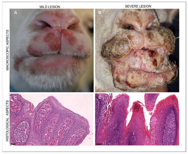
Figure 2.
Representative images of the macroscopic appearance of ORFV-infected lambs and microscopical patterns of ORFV-infected lambs with mild and severe lesions. (A) Mild lesions in an ORFV-infected lamb showing vesicles and crusted pustules in the upper and lower lips. (B) Severe lesions appearing in an ORFV-affected lamb as coalescing hyperemic, proliferative, verrucous outgrowths with a papilloma-like appearance covering the incisor tooth, present in the lips together with crusted lesions. (C) Mild lesions histologically showing moderate epithelial hyperplasia, hyperkeratosis with elongated rete ridges, and proliferations of neovascular structures in the dermis of the host skin. Hematoxylin and eosin staining (H&E). Original magnification 100×. Scale bar = 100 µm. (D) Severe manifestation of ORFV-infected animals microscopically characterized by massive proliferative patterns involving the epithelium and showing hyperkeratosis and hyperplastic epithelium with extremely elongated rete ridges and ballooning degeneration. In the lamina propria of the buccal mucosae and in the derma of skin, there is evident proliferation of mesenchymal cells resembling hemangiomatous patterns. Hematoxylin and eosin staining (H&E). Original magnification 100×. Scale bar = 100 µm.

